Top-tip: Supraventricular TachycardiaArticle 10 of 13
Tachycardias
Tachycardias are the most exciting part of learning about ECGs. It is of course important to understand the mechanism causing tachycardias, but it is equally important to remember to look at the patient first. "Benign" arrhythmias can compromise a person, and "serious" arrhythmias can be asymptomatic. Look at the patient and act with commonsense, rather than treating the ECG.
We are not going to try to explain the emergency treatment of arrhythmias, but this should be something on all doctors' minds (and hearts). All the emergency treatment protocols are available from the European Resuscitation Council (website www.erc.edu/).
Tachyarrhythmias can be divided into two broad categories: supraventricular tachycardias (SVT) and ventricular tachycardias (VT). It is useful to think of these as narrow complex (QRS complex <120ms) tachycardias (SVT) and broad complex (QRS >120 ms) tachycardias (VT).
Why is the QRS complex narrow?
As described above, normally the atrium passes the sinoatrial (SA) node's signal to the atrioventricular (AV) node. This then passes on through the ventricular specialised conducting tissue, which gives rapid, synchronous activation of the whole of the ventricle. Thus to get a narrow complex you must have an electrical signal that passes forward through the AV node, and you must have specialised conducting tissue, that works. A broad QRS complex means that either your conducting system is not working (bundle branch block) or the electrical circuit is not involving the AV node correctly.
So what causes arrhythmias?
Only two things cause abnormal heart rhythms. The most common mechanism involves an abnormal electrical circuit which allows the heart beat to cycle around it. This is called re-entry. The other is where a focal area of the heart starts to "spark off" and send out a shower of extra heart beats (increased automaticity or triggered activity; see figure 1).
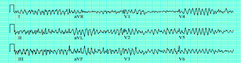
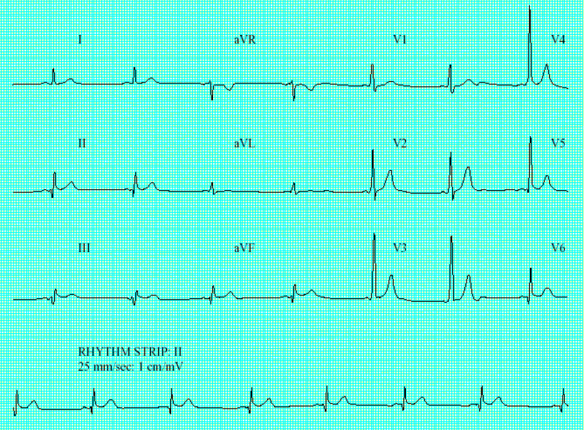
ECG 1: Atrial fibrillation.

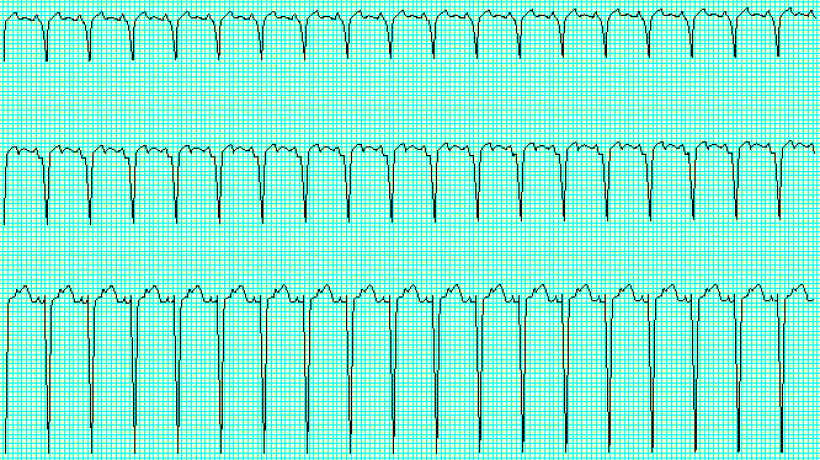
ECG 2: Atrial flutter.
Narrow complex tachycardias
Atrial fibrillation (AF):
- The most common sustained arrhythmia of all
- Affects 2% of 65-75 year-olds, 5% over 75 years
- Associated with any disease affecting the heart
- Other narrow complex tachycardias can degenerate to AF--for example, atrial flutter
- Re-entrant mechanism.
Arrhythmia anatomy: Multiple wavelets of re-entry swashing around the atria. The AV node is inundated with cascades of chaotic activity (see figure 2).
ECG: Irregular ventricular rate with no true P waves, baseline irregularities representing atrial activation (ECG 1).
Useful drugs: anticoagulation to prevent thromboembolism; ß blockers, sotalol, propafenone, flecainide, and amiodarone to prevent paroxysmal AF; digoxin ß blocker, diltiazem, and verapamil (often in selected combinations) to control heart rate.
Atrial flutter:
- Mistakenly thought of as a variant of atrial fibrillation
- Occurs at any age
- Can be difficult to control with drug therapy
- Re-entrant mechanism.
Arrhythmia anatomy: Typically involves a large circuit created by the structures in the right atrium (figure 3).
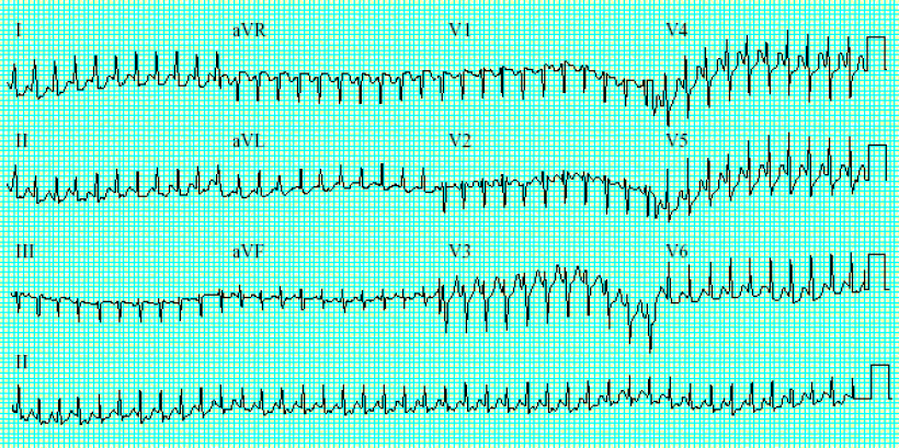
ECG 3: AV node re-entrant tachycardia during tachycardia.
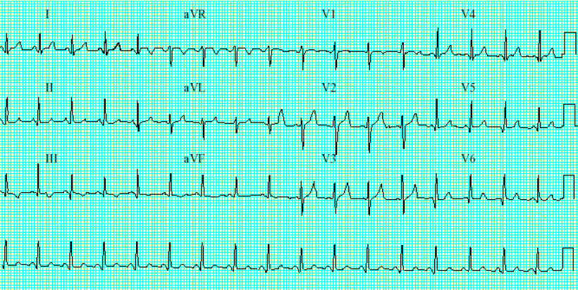
ECG 4: The same patient with AV node re-entrant tachycardia now in sinus rhythm. The QRS complex in lead V1 no longer has a second small R wave, which was due to backward atrial depolarisation occurring at the same time as the QRS during palpitations.
ECG: Saw tooth baseline with atrial rate of 300 and ventricular response of 150. Always be suspicious of flutter if you see a narrow QRS tachycardia at a heart rate of 150(ECG 2). Flutter waves are seen before each QRS and after each QRS in the middle of each T wave.
Useful drugs: Digoxin and verapamil (or diltiazem) can be used to slow the response of the AV node. You still get an atrial rate of 300 but hopefully a slower ventricular rate. Type 1c agents (propafenone or flecainide) can be used to stop the atrial circuit and terminate the tachycardia.
Atrioventricular nodal re-entry tachycardia (AVNRT)
- Also known as AV nodal tachycardia (AVNT), or AV junctional tachycardia (AVJRT)
- Commonest type of narrow QRS
- Typically starts in the later teens and 20s
- Re-entrant mechanism.
Arrhythmia anatomy: The basis of this arrhythmia is that the AV node can have two pathways as part of it (see figure 3). These paths allow a circuit to be set up at the AV node itself, giving rise to the arrhythmia.
ECG: Narrow QRS tachycardia of about 180 bpm with P waves often absent: they are hidden in the QRS, as atrial and ventricular depolarisation occur at the same time. You may see changes in the QRS complex in lead V1 as compared with the resting ECG: these are due to simultaneous P wave activation. This is a small positive deflection before the onset of the T wave and is shown in ECG 3 and 4. Look at the subtle difference in the QRS during tachycardia and during normal rhythm.
Useful drugs: Drugs which affect the AV node--that is, digoxin, diltiazem, and verapamil; adenosine can acutely stop the tachycardia as it transiently blocks the AV node (as can vagotonic manoeuvres); ß blockers; type 1c agents (propafenone or flecainide).
Atrial tachycardias
- Second most common SVT
- Can occur at any age, but increased likelihood if the atria are diseased (hypertension, pulmonary disease, previous cardiac surgery, etc)
- Either focal automatic activity (mainly) or due to re-entrant mechanism. Can have multiple foci of automatic activity.
Arrhythmia anatomy: Varied anatomical basis (see figure 4): they can be due to a focal area in the atria with increased automaticity, or re-entry around an abnormal area of atrial tissue.
ECG: Abnormal looking P waves; may have different types of P waves on same ECG. Heart rate very variable from 140 to 240 bpm.
Useful drugs: ß blockers; verapamil or diltiazem; type 1c agents (propafenone or flecainide); amiodarone.
Atrioventricular re-entrant tachycardia (AVRT) and Wolff-Parkinson-White syndrome
- Can occur from infancy onwards
- In infants it may be associated with congenital heart defects
- Re-entrant mechanism
- One to three people in every 1000 have an obvious extra pathway on the resting ECG--that is, Wolff Parkinson White syndrome. It may even be you!
Arrhythmia anatomy: The re-entry mechanism is due to a congenital addition of a small piece of atrial tissue which crosses the isolating fibrous ring separating the atria and ventricles. This gives a large circuit with the heart beat passing down through the AV node, around the ventricle, back up the pathway, and across the atrium back to the AV node. This illustrates why SVT is not such a good name for these arrhythmias: most of this tachycardia's circuit lies in the ventricle.
ECG: The resting ECG can be normal but it can show evidence of the pathway's existence if the path allows some of the atrial depolarisation to pass quickly to the ventricle before it gets though the AV node (Wolff-Parkinson-White (WPW) syndrome; ECG 5). The early depolarisation of part of the ventricle leads to a shortened PR interval and a slurred start to the QRS (delta wave). The QRS is narrow; the message via the AV node eventually predominates because it uses the rapid conducting system to depolarise most of the ventricle.
The tachycardia ECG may be unremarkable, with P waves absent (hidden in the QRS). If the circuit is long or slow enough the P wave may occur at the end of the QRS and can be visible as a distortion in the T wave (this is best seen in lead V1).
Useful drugs: ß blockers; adenosine can acutely stop the tachcardia as it transiently blocks the AV node (as can vagotonic manoeuvres); type 1c agents (oropafenone or flecainide); type 1a agents (procainamide or quinidine).
Atrial fibrillation has an atrial rate of 300-600 bpm. Fortunately, the AV node protects the ventricle from experiencing such a heart rate. In patients with accessory paths there is a mechanism for this very fast heart rate to bypass the AV node and cause AF with a dangerously fast ventricular response (ECG 1). Treating this with drugs such as digoxin, verapamil, or diltiazem would further block the AV node but not prevent the AF from passing down the accessory path. The situation would be made a lot worse in this case. This is why the drugs are stated as being contraindicated in atrial fibrillation in the presence of an accessory pathway.
ECG 5: Wolff-Parkinson-White syndrome.
ECG 6: A patient with AF and an accessory pathway. The ECG is irregular and frighteningly quick (more than 300bpm in places).
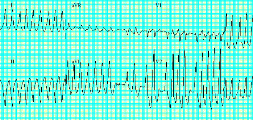
ECG 7: Ventricular tachycardia: AV dissociation.
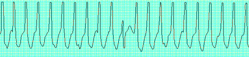
ECG 8: Ventricular fibrillation.