Top tips: Chest X-Rays - Step 3, Heart contoursArticle 15 of 21
The heart size can only be assessed confidently on a PA X-ray. The heart is considered enlarged if the Cardio-Thoracic ratio (CTR) is >50%.
Figure 30: CTR as measured by the widest transverse diameter of the heart and widest transverse diameter of the thoracic cavity.
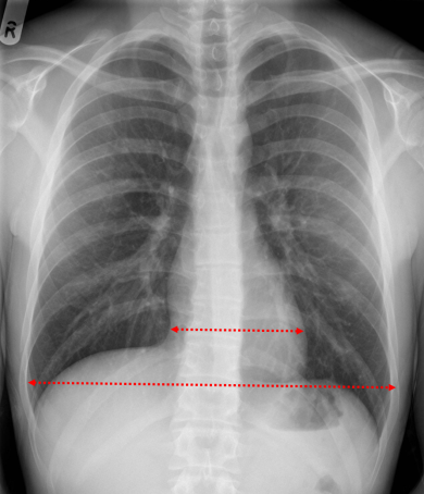
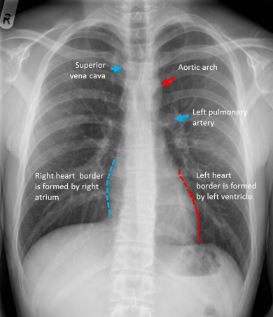
Figure 31: The right and left heart borders and borders of the mediastinum
The most common reason for the heart to be enlarged is congestive cardiac failure, so look for signs of left ventricular failure on the rest of the film. These are:
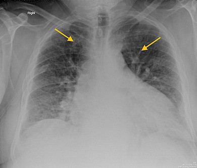
- Upper lobe blood diversion- The pulmonary veins running from the upper lobes seem more prominent than those running from the lower lobes.
- Kerley B lines- these are tiny horizontal lines from the pleural edge and are typical of fluid overload with fluid collecting in the interstitial space.
- "Bat's wing" haziness around the hila.
- Alveolar shadowing-in very severe pulmonary oedema fluid collects not only in the interstitial space but in the air spaces or alveoli. You can recognise this by seeing hazy shadowing throughout the lungs, and possibly air bronchograms.
There are only a few occasions when there may be the appearance of left ventricular failure (LVF) but a normal sized heart - in an acute myocardial infarct (sudden onset of LVF). Lymphangitis carcinomatosa may mimic the appearances of LVF and be accompanied by a normal sized heart.
Figure 32: Cardiomegaly
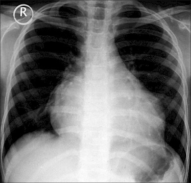
Figure 33: There is mild cardiomegaly with bilateral small pleural effusion (blunting of both costophrenic angle) consistent with features of heart failure. Note also the presence of median sternotomy.
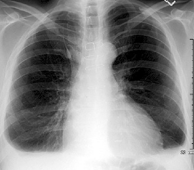
Figure 34: Example of a patient presenting acutely with pulmonary oedema. There is bilateral perihilar 'bat wing like' opacification and small basal pleural effusion.
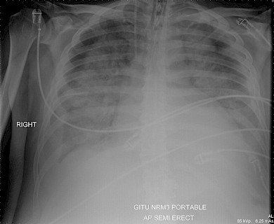
Figure 35: This is an example of heart failure with presence of cardiomegaly, upper lobe venous diversion (arrows) and subtle septal lines in the bases.