Top tips: Chest X-Rays - Step 3, Lung changesArticle 19 of 21
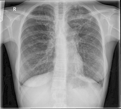
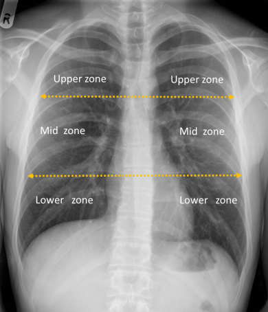
The lungs are divided into zones and when describing chest X ray use the term ‘zones’ instead of lobes. They are divided as upper (above the hilum), middle and lower zones (below the hilum).
Figure 46: Chest showing lung zones
The lung is made up of bronchi, which branch, at the end of which are alveoli. The interstitial space is the potential space which surrounds the alveoli. The whole of the lung from bronchi to alveoli is the air space - that is, it normally contains air. But the air spaces can fill up - with fluid (such as in pulmonary oedema), with pus (as in infection), with blood (as in rare diseases that causes alveolar haemorrhage), or with tumour cells (adenocarcinoma). Fluid and pus are by far the commonest. When the air spaces fill up, the alveoli fill first, with the bronchi being relatively spared. Therefore the bronchi, which are still air filled, stand out against the alveoli, which are filled with pus or fluid. This is called an ‘air bronchogram’ and is simply a sign that there is air space disease. Consolidation is another term for airspace shadowing.
If there is air space disease then you need to work out which part of the lungs it is affecting. Something like "there is shadowing in the air spaces of the right mid and lower zone” is a simple way to describe the changes.
Single nodule or mass in the lung: The two main causes of a single mass in the lung are infection and neoplasm.
Figure 47: There is right upper lobe consolidation with presence of air-bronchogram (arrow).
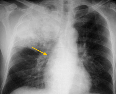
Figure 48: There is large mass in the left upper lobe which is consistent with a lung cancer.
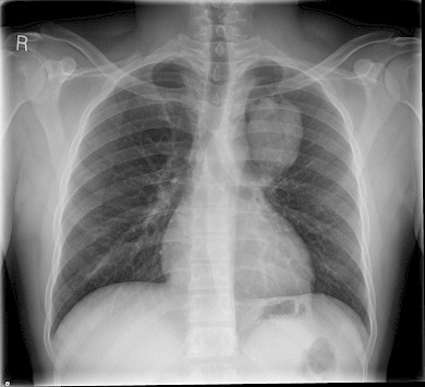
Figure 49: The right hilum is bulky secondary to a right hilar mass (arrow) which represents a lung cancer.
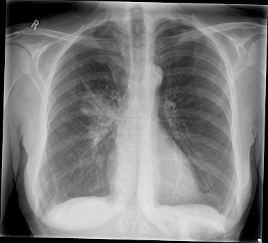
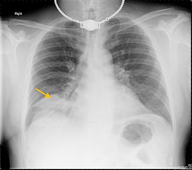
Figure 50: Right middle lobe consolidation in a patient with infection (arrow). Also note the presence of tracheostomy.
Differential diagnosis of small and large lung nodules Small nodules (<5 mm)
- Common: Metastases (especially breast, testis, gastrointestinal tract, kidney, and thyroid)
- Less common: Miliary tuberculosis (so called because they look like tiny seeds or "milia" and are due to haematogenous spread)
- Sarcoid (which can also cause reticulo-nodular shadowing)
- Pneumoconiosis - for example, due to inhaling coal dust. This is rare nowadays
- Chickenpox pneumonia Larger nodules/masses (>5 mm)
- Rare: Inflammatory nodules - for example, due to vasculitis of rheumatoid arthritis or Wegener's granulomatosis
Figure 51: Bilateral lung nodules in a patient with metastatic lung disease.
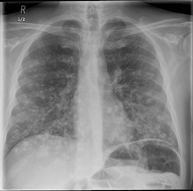
Figure 52: Multiple 'miliary' nodules in a patient with miliary TB
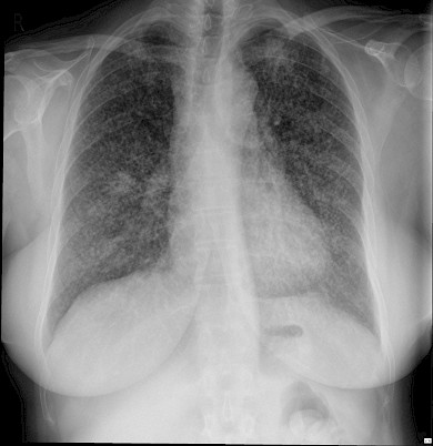
Figure 53: Extensive multiple 'cannon ball' lung nodules consistent with lung metastasis in a patient with known sarcoma.
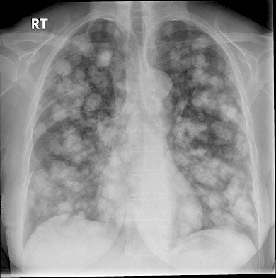
Figure 54: A patient with pulmonary sarcoidosis showing bilateral hilar lung adenopathy and subtle bilateral lung nodules.
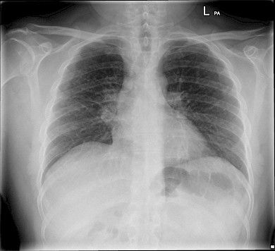
Differential diagnosis of cavitating lung lesions
- Common: infection bacterial infection such as staphylococcus
- Tuberculosis, look out for mycetoma is a pre-exiting cavity
- Cavitating malignancy
- Rare: septic embolic
- vasculitis
Figure 55: There is a cavity in the right upper lobe. Notice the presence of a soft tissue density (arrow) with a presence of 'air crescent' around it consistent with a fungal ball or mycetoma.
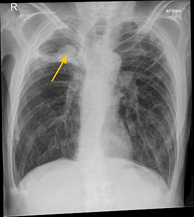
Figure 56: There is consolidation with cavitation in the right upper lobe in a patient with staphylococcus infection.
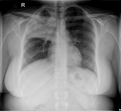
Figure 57: Multiple cavitating lung lesion with 'air fluid' in a patient with septic embolic.
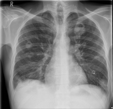
Next let’s look at the interstitial space. This surrounds bronchi, vessels, and groups of alveoli. When there is disease in the interstitium it manifests itself by reticulo-nodular shadowing (crisscross lines or tiny nodules or both). The main two processes affecting the interstitium are accumulation of fluid (occurring in pulmonary oedema or in lymphangitis carcinomatosa) and inflammation leading to fibrosis. If you see crisscross lines or tiny nodules or both say: "There is reticulo-nodular shadowing within the lower zones."
Figure 58: Pulmonary fibrosis with volume loss and bilateral increased interstitial lung marking.
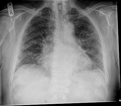
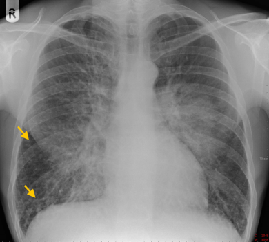
Figure 59: There is bilateral opacification around the hilar region with peripheral multiple septal lines (arrows). There are features of acute pulmonary oedema.
Complete opacification of the hemi-thorax
There are four main causes of opacification of a hemi-thorax –
- Consolidation (that is, material within the air-spaces
- Pleural effusion - that is, material within the pleural space, which could be serous fluid, blood, or pus
- Complete collapse of one lung with the mediastinum shifting over the abnormal side can also cause a "white out" on the abnormal side.
- Finally, after a pneumonectomy the mediastinum shifts to the empty hemithorax and the residual pleural space fills with fluid and fibrotic material leaving the patient with a complete "white out" on the side that has been operated on.
Consolidation and pleural effusion are the two most common, and it can be difficult to distinguish between them - of course, they can coexist.
Finally summarise your findings as you are looking: "The trachea is central, the mediastinum is not displaced. The mediastinal contours and hila seem normal. The lungs seem clear, with no pneumothorax. There is no free air under the diaphragm. The bones and soft tissues seem normal."
If you have not seen any abnormality by this point, say so - "I have not yet identified an abnormality so I will now look through my review areas" - and then look at the "review areas" - places where you can easily miss disease.
Figure 60: Left pneumonectomy.
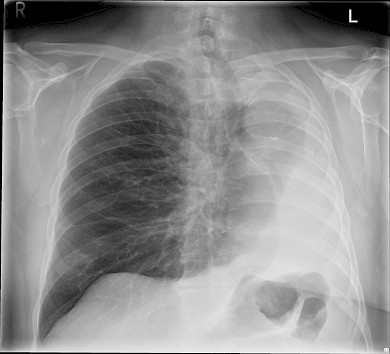
Figure 61: This opaque hemithorax is caused by complete left lung collapse. Note the presence of mediastinal and tracheal shift to the left.
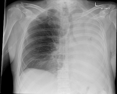
Figure 62: Example of bronchiectasis with bilateral ' ring shaped' areas representing dilated thick wall bronchus seen 'end on'.