Top tips: Chest X-Rays - Step 3, Lung lobes & fissuresArticle 11 of 21
Now look at the lungs. Apart from the pulmonary vessels (arteries and veins), they should be black (because they are full of air). Scan both lungs, starting at the apices and working down, comparing left with right at the same level, just as you would when listening to the chest with your stethoscope. The lungs extend behind the heart, so look here too. Force your eye to look at the periphery of the lungs - you should not see many lung markings here; if you do then there may be disease of the air spaces or interstitium.
The left lung has two lobes and the right has three lobes and these have separate pleural coverings. There is an oblique fissure and a horizontal fissure on the right and an oblique fissure on the left separating the lung into lobe. This lobar anatomy is essential in identifying the lobe that is collapsed.
Lobar Collapse:
When the lobe is not aerated it will lose much of its volume and collapse to a predictable location depending on whether it is an upper, middle, or lower lobe. The collapsed lobe itself can be very difficult to see - there may simply be a little extra shadowing on the film.
The signs that should alert you to collapse due to the loss of lung volume are:
- The mediastinum may be shifted towards the side of collapse
- The hilum is pulled up or down from where it normally lies
- The horizontal fissure will also be pulled up or down
- The remaining (non-collapsed) lung on the side of the collapse has to expand to fill the hemi-thorax, thus "spreading" it’s contained vessels; therefore the abnormal side will seem blacker with fewer lung markings than the opposite normal side
Collapse of a lobe is caused by proximal obstruction - for example, by a neoplasm, mucus plug (such as in a postoperative patient), or foreign body (such as in a child). Always mention that you are looking for the cause of the collapse. Check if the proximal obstruction is visible - for example, a large carcinoma arising from the right upper lobe.
Right upper lobe collapse:
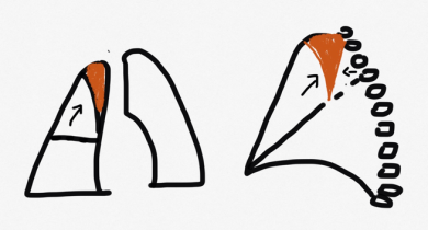
Figure 13: Right upper lobe collapse. There is volume loss with the right hilum being pulled up and trachea displaced to the right.
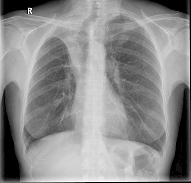
- Trachea deviated to the right
- Horizontal fissure & right hilum pulled up
- Triangular opacity on lateral x ray.
Right middle lobe collapse:
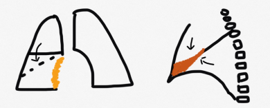
Figure 14: Right middle lobe collapse
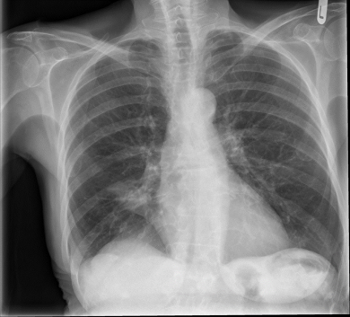
- Loss of right heart border
- Ill-defined opacity adjacent to right heart border
- Horizontal fissure pulled downwards
- Triangular opacity on lateral X ray
Right lower lobe collapse:
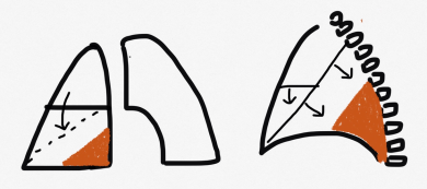
Figure 15: This is an example of a right lower lobe collapse with the increased density adjacent to the right heart border.
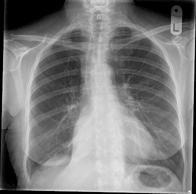
- Well defined opacity adjacent to the right heart border
- Oblique and horizontal fissures displaced posteriorly
Left upper lobe collapse:
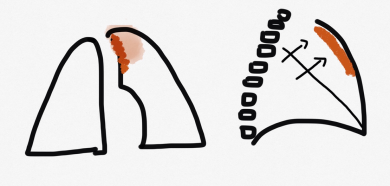
Figure 16: Left upper lobe collapse with 'veil' like opacity in the left upper lobe. Not also the presence of a small left basal pleural effusion.
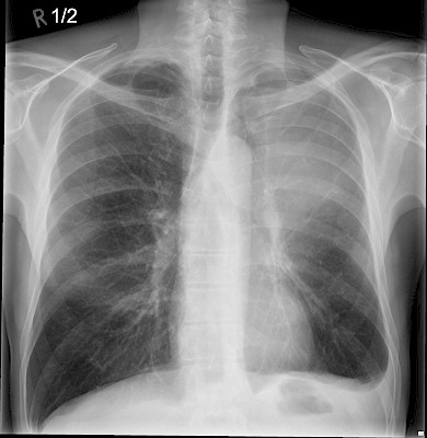
- Trachea pulled to the left
- Ill-defined opacity/veil like opacity
- Left hilum pulled upwards
Left lower lobe collapse:
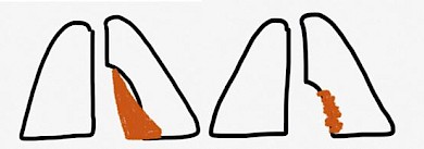
Figure 17: This X ray show a tight left lower lobe collapse with increased ‘sail-like’ density (arrow) behind the left heart border.
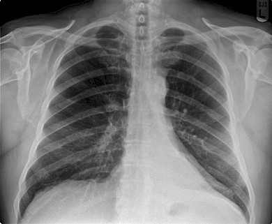
- Left lower lobe collapse- Triangular opacity behind the heart with loss of medial border of diaphragm
- Lingular collapse – ill-defined left heart border.